Hip Imaging and Investigations
Imaging is necessary to confirm a diagnosis, evaluate the extent of the condition and rule out other associated conditions. It helps to formulate and plan both non-operative and operative management.
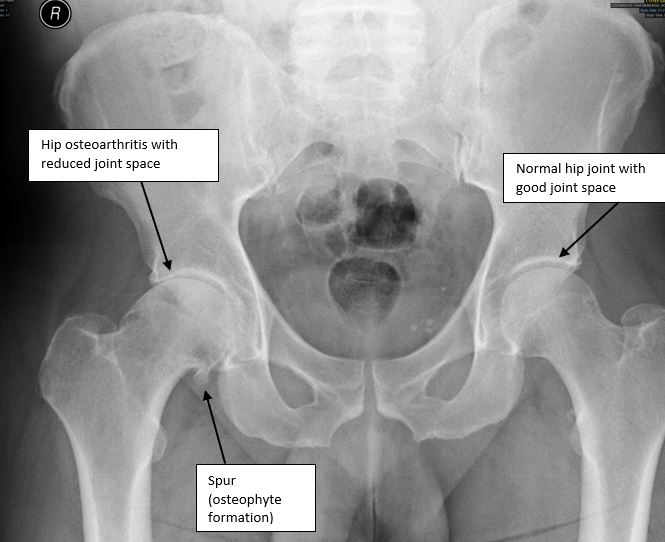
Plain radiographs (X-rays) are usually the first line of investigation to understand basic bony anatomy and to confirm diagnosis (Fig 1). The majority of cases such as osteoarthritis, hip impingement, developmental abnormalities, like hip dysplasia, and other conditions can be confirmed and assessed with X-rays.

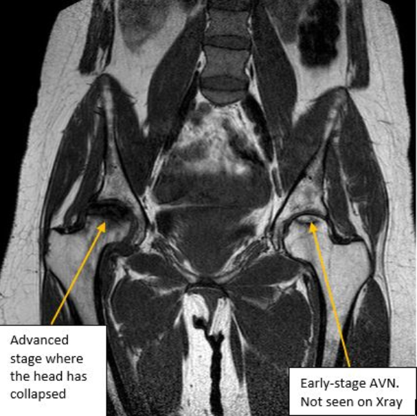
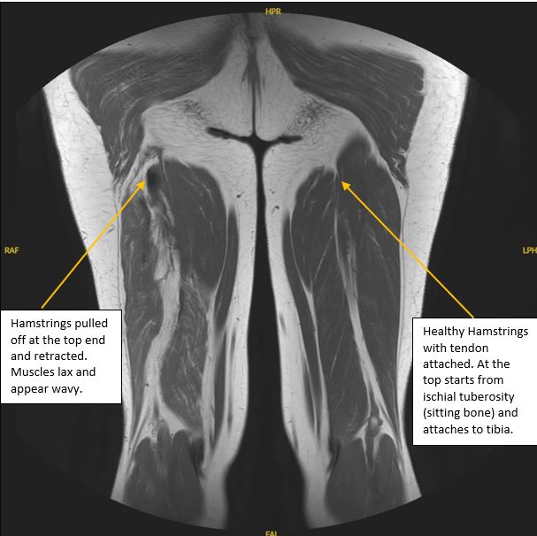
Magnetic Resonance Imaging (MRI) shows more clearly soft tissues such as labrum, ligaments, muscles and tendons around the hip joint. Sometimes it helps in assessing the joint cartilage and changes in the adjacent bone which helps diagnose conditions such as localized hip osteoarthritis, osteochondral defects, etc. Labral changes (changes to the fibro cartilage – one of the three types of cartilages in our body) are commonly found in patients after a certain age and also in patients with hypermobile joints, but these changes need to be correlated with physical and other important investigation findings, such as X-rays and CT scans, before confirming hip impingement. MRI is accurate in diagnosing tendon injuries and their severity like hamstrings traumatic rupture (Fig 2). It is also the investigation tool used to diagnose avascular necrosis (AVN) of the hip in its early stages (Fig 3).


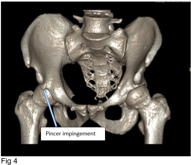
Computerised Tomography (CT) scan is the preferred investigative tool to view the hip bone anatomy in three dimensions (3D). This helps to diagnose hip conditions not easily seen on X-rays, for example hip impingement and to plan treatment accordingly (Fig 4). It is a very important diagnostic tool in assessing failed hip replacements, bone defects and alignment of prostheses in situ. CT or MRI are also used to plan robotic or computer-navigated hip replacements and other orthopaedic procedures.

Occasionally alternative imaging techniques, such as a bone scan, are necessary to investigate the hip problem as this type of imaging is helpful in identifying infection, loose prostheses, tumours, etc. It is very useful in patients who cannot have MRI due to various medical reasons. Additionally, blood investigations are essential in assessing inflammatory, infection and metabolic conditions affecting the hip joint.
